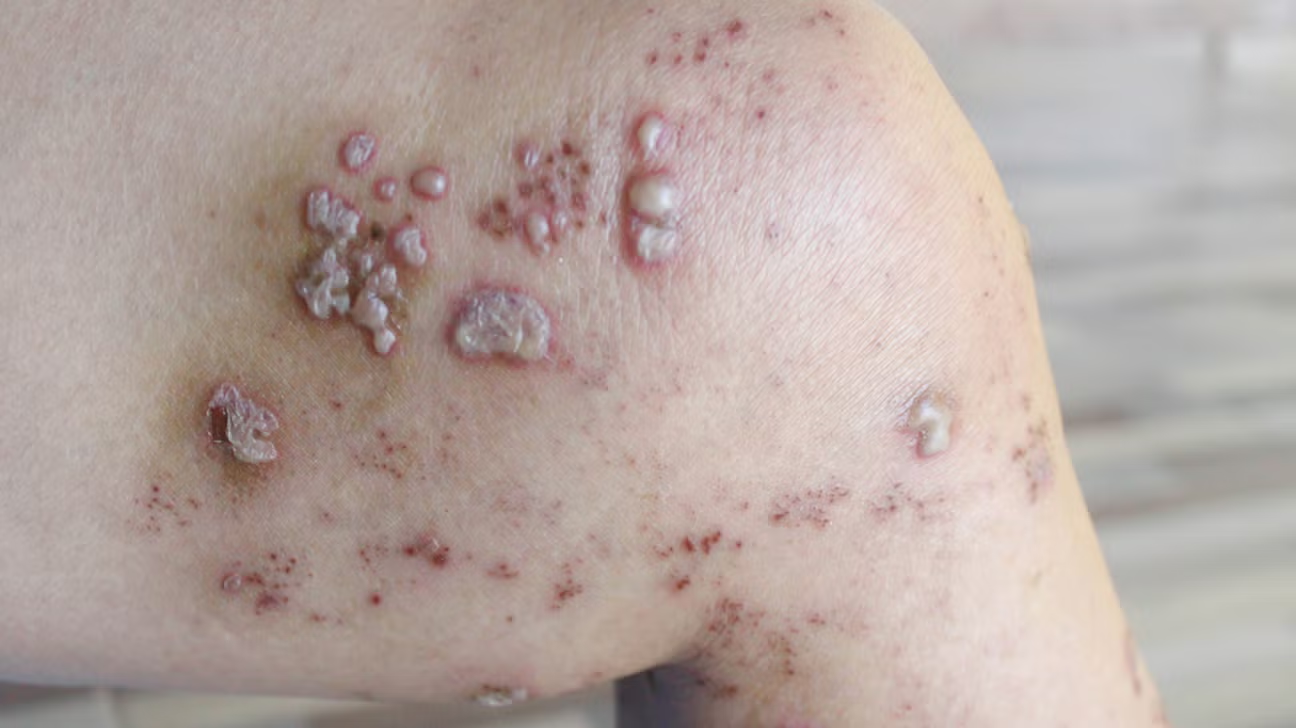
The general practice setting functions as the frontline for an immense array of dermatological issues, presenting a daily challenge to clinicians who must rapidly and accurately interpret the visual language of the skin, a skill often underrepresented in foundational medical training. Skin conditions are remarkably common, representing a significant portion of patient visits, yet the sheer variety and the subtle morphological distinctions between benign, inflammatory, and malignant processes can often lead to diagnostic uncertainty. The stakes are particularly high when attempting to discern between self-limiting eruptions and those demanding urgent specialist referral or systemic intervention. Navigating this clinical landscape necessitates a systematic approach that moves beyond mere pattern recognition, delving into the patient’s comprehensive history, exploring genetic predispositions, and meticulously noting the specific topography and characteristic features of the cutaneous lesions. The lack of direct visual training and the reliance on textbooks that often disproportionately feature lighter skin tones compound the difficulty, making the application of classic textbook descriptions a frequent source of error in diverse patient populations. It is precisely at this juncture—the interface between common, self-manageable conditions and those with serious implications—that the expertise and clinical acumen of the primary care provider are most rigorously tested. This is a field where misinterpretation of a single lesion can have disproportionate health consequences, underscoring the necessity for continual refinement of diagnostic skills and an open pathway to expert consultation.
The sheer variety and the subtle morphological distinctions between benign, inflammatory, and malignant processes can often lead to diagnostic uncertainty
The core difficulty in primary care dermatology stems from the visual overlap among vastly different disease etiologies. “The sheer variety and the subtle morphological distinctions between benign, inflammatory, and malignant processes can often lead to diagnostic uncertainty” encapsulates this fundamental challenge. A red, scaly patch, for instance, could be an innocuous case of tinea corporis (ringworm), a flare of chronic plaque psoriasis, or, less commonly but more critically, a manifestation of cutaneous T-cell lymphoma. Without adequate experience and a systematic method of examination, a primary care physician risks initiating an inappropriate treatment path—treating a fungal infection with steroids, for example, which can dramatically worsen the condition—or, conversely, delaying the timely diagnosis of an emerging skin malignancy like basal cell or squamous cell carcinoma. The presentation of common conditions such as eczema and psoriasis can also shift based on anatomical location, age of the patient, and skin phototype, further obscuring the “classic” findings described in most educational materials.
The presentation of common conditions such as eczema and psoriasis can also shift based on anatomical location, age of the patient, and skin phototype
Understanding how common inflammatory dermatoses deviate from their textbook presentation is vital for accurate triage and management in a generalist setting. “The presentation of common conditions such as eczema and psoriasis can also shift based on anatomical location, age of the patient, and skin phototype” highlights the variability inherent in these widespread conditions. Atopic dermatitis, or eczema, typically presents in the creases of joints (antecubital and popliteal fossae) in older children and adults, yet infants commonly show involvement on the face and extensors. Similarly, while classic plaque psoriasis is defined by well-demarcated, thick, silvery-scaled plaques on the elbows and knees, inverse psoriasis, found in skin folds, lacks the scale and may be mistaken for a fungal or candidal infection. In individuals with richly pigmented skin, both conditions can lack the vivid erythema often described, instead manifesting as violaceous, brown, or grey patches, making the visual diagnosis far more challenging without a high index of suspicion.
In individuals with richly pigmented skin, both conditions can lack the vivid erythema often described, instead manifesting as violaceous, brown, or grey patches
The impact of skin color on the clinical presentation of dermatological disease is a critical, yet frequently overlooked, area of diagnostic skill development for non-dermatologists. “In individuals with richly pigmented skin, both conditions can lack the vivid erythema often described, instead manifesting as violaceous, brown, or grey patches” speaks directly to the limitations of educational resources predominantly illustrating diseases on lighter skin. A provider relying solely on the presence of bright redness to indicate inflammation will consistently under-diagnose or mischaracterize conditions in patients of color. Furthermore, post-inflammatory hyperpigmentation—a dark discoloration left after an inflammatory lesion resolves—is far more pronounced and persistent in darker skin tones, often leading to patient distress and therapeutic interventions aimed at the wrong target. Recognizing subtle color changes and relying more heavily on palpation (for texture, induration, and scale) and pattern recognition becomes essential to compensate for the loss of the diagnostic cue provided by clear erythema.
Recognizing subtle color changes and relying more heavily on palpation (for texture, induration, and scale) and pattern recognition becomes essential
Moving beyond color as the sole indicator necessitates a more tactile and analytical approach to the skin examination, integrating information from texture and distribution. “Recognizing subtle color changes and relying more heavily on palpation (for texture, induration, and scale) and pattern recognition becomes essential” suggests a framework for refining the clinical eye. For example, in acne vulgaris, the primary care provider must distinguish between non-inflammatory comedones and inflammatory papules or nodules, guiding the choice between topical retinoids, benzoyl peroxide, or oral antibiotics. Palpation helps to define the depth of the lesion—a deep, tender nodule suggesting a need for systemic therapy, versus superficial pustules amenable to topical treatment. The concept of pattern recognition also extends to common viral exanthems, like Pityriasis Rosea, where the initial ‘herald patch’ followed by a ‘Christmas tree’ distribution along the cleavage lines of the back provides a classic, though not always present, diagnostic clue that can rule out conditions like secondary syphilis or tinea corporis.
The concept of pattern recognition also extends to common viral exanthems
The ability to recognize specific, non-inflammatory patterns allows the primary care physician to confidently reassure patients and avoid unnecessary treatment. “The concept of pattern recognition also extends to common viral exanthems” illustrates the utility of distributional cues in diagnosing self-limiting conditions. A prominent example is the widespread occurrence of various tinea infections, collectively known as dermatophytoses, such as Tinea Corporis (ringworm). While the classic lesion is an annular patch with central clearing and an active, scaly border, variations are routine. A correct diagnosis, which can often be confirmed rapidly by a potassium hydroxide (KOH) preparation in the office, dictates the use of topical antifungals, avoiding the clinical trap of mistaking it for eczema or psoriasis and applying topical steroids, which would only feed the fungus and cause an exaggerated, or ‘tinea incognito,’ presentation.
A correct diagnosis, which can often be confirmed rapidly by a potassium hydroxide (KOH) preparation in the office
The availability of simple, in-office diagnostic procedures significantly improves the accuracy of primary care dermatology, offering rapid confirmation for common infectious etiologies. “A correct diagnosis, which can often be confirmed rapidly by a potassium hydroxide (KOH) preparation in the office” underscores the value of accessible diagnostic tools. The KOH prep, which dissolves keratin and allows for visualization of fungal hyphae, is an indispensable asset for definitively diagnosing tinea infections and distinguishing them from non-infectious conditions like nummular dermatitis. Similarly, performing a brief scraping for scabies mites or using a dermatoscope to examine pigmented lesions allows for a level of diagnostic precision that far exceeds simple naked-eye observation. Equipping and training primary care staff to confidently perform these few, high-yield procedures can dramatically reduce unnecessary specialist referrals and expedite effective patient care for highly prevalent conditions.
Equipping and training primary care staff to confidently perform these few, high-yield procedures can dramatically reduce unnecessary specialist referrals
Strategic deployment of simple diagnostics enables the primary care setting to efficiently manage the vast majority of common skin complaints without specialist intervention. “Equipping and training primary care staff to confidently perform these few, high-yield procedures can dramatically reduce unnecessary specialist referrals” points to a clear quality improvement metric. Consider the diagnosis of verrucae (warts) caused by the Human Papillomavirus (HPV). While visually recognizable, differentiating warts from common benign lesions like seborrheic keratoses or molluscum contagiosum can sometimes be subtle. The use of dermoscopy aids in visualizing the characteristic thrombosed capillaries (black dots) within the warty surface. For other benign, but often cosmetically bothersome, lesions such as seborrheic keratoses, which have a “stuck-on” or “waxy” appearance, reassurance to the patient of their benign nature is often the only required action, saving specialist time for more complex, life-threatening pathologies.
For other benign, but often cosmetically bothersome, lesions such as seborrheic keratoses, which have a “stuck-on” or “waxy” appearance
The primary care provider also plays a crucial role in distinguishing between various types of benign growths and potentially malignant lesions, particularly when addressing patient anxiety. “For other benign, but often cosmetically bothersome, lesions such as seborrheic keratoses, which have a “stuck-on” or “waxy” appearance” is a classic visual descriptor that helps categorize these extremely common lesions. However, the essential skill in the primary care setting remains the effective screening for skin cancer, specifically the use of the ABCDE criteria (Asymmetry, Border irregularity, Color variation, Diameter greater than 6mm, and Evolving change) for melanoma. Teaching patients to perform regular self-examinations and maintaining a low threshold for suspicious lesions—especially those that are non-uniform or have changed rapidly—is a critical public health function performed daily by general practitioners, representing a major protective factor against advanced malignancy.
Teaching patients to perform regular self-examinations and maintaining a low threshold for suspicious lesions—especially those that are non-uniform or have changed rapidly
Empowering patients to be vigilant about changes to their skin acts as an essential, decentralized surveillance system for early detection of malignancy. “Teaching patients to perform regular self-examinations and maintaining a low threshold for suspicious lesions—especially those that are non-uniform or have changed rapidly” emphasizes the collaboration required between clinician and patient in skin cancer prevention. The clinician’s role extends to managing chronic, recurring conditions such as urticaria (hives). While typically a transient allergic reaction, chronic urticaria lasting longer than six weeks often signals an underlying autoimmune or systemic issue. A thorough history is paramount, exploring recent exposures, medications, and systemic symptoms before initiating first-line therapy with H1 antihistamines. This investigative step is a classic example of where the generalist’s broad perspective is crucial, linking a seemingly isolated skin problem to a potential internal pathology that requires deeper exploration.
A thorough history is paramount, exploring recent exposures, medications, and systemic symptoms before initiating first-line therapy with H1 antihistamines
The final step in successful primary care dermatology is the synthesis of physical findings with a detailed patient narrative to formulate a comprehensive management plan. “A thorough history is paramount, exploring recent exposures, medications, and systemic symptoms before initiating first-line therapy with H1 antihistamines” underscores the necessary transition from visual diagnosis to contextual management. The primary care provider must master the use of topical steroids—knowing when to use a low-potency cream versus a high-potency ointment, and for how long—and understand the contraindications for common systemic agents, such as avoiding tetracycline antibiotics for acne in pregnancy. Ultimately, the competence of a general practitioner in dermatology is measured not by their ability to treat the esoteric, but by their consistency in correctly managing the common, confidently identifying the truly malignant, and knowing the precise moment to transfer care to a specialist for complex or undiagnosed conditions.